Creative Proteomics Announces the Launch of New Website in Tumor Multi-Omics

Creative Proteomics is very excited to announce the launch of its newly designed website. After years of hard work and dedication, Creative Proteomics is delighted to officially announce it. Creative Proteomics Tumor Multi-Omics, as the name of new website, fully integrates the company’s core technologies in solutions for cancer researches, including multi-omics services, i.e. (meta) genomics, (meta) transcriptomics, (meta) proteomics and metabolomics. The company’s success is based on its excellent R & D, technology, and service teams, as well as its internationally leading technology platforms and high-tech.

As a leader in oncology field, Creative Proteomics endeavors to provide researchers with the most effective solutions, whether cancer studies starting at the genomic level, elucidating cellular pathways, determining the effects of compounds on 3D tumor models, studying tumor growth in animal models, or studying tumors and their microenvironment.

The goal with this new website is to provide scientists an easier way to explore based on their own choice. The new website gives better access to What the Company Offers:

Proteomics Service: Protein Gel and Imaging, Protein ldentification, Protein Quantification, Protein Post, Translational Modification Analysis, Top Down Proteomics, Peptidomics Analysis, Protein-Protein Interaction Networks, Subcellular Proteomics, and so on.

Metabolomics Service: Untargeted Metabolomics, Targeted Metabolomics, Metabolic Flux Analysis (MFA), Unknown Metabolites ldentification, Xenobiotic Metabolites Analysis, Untargeted Lipidomics, Targeted Lipidomics, Exosome Llpidomics, MALDI-Imaging Lipidomics.

Transcriptomics Service: Gene Chip Technology Services, RNA Sequencing, LC-MS, tRNA Sequencing, tRF&tiRNA Sequencing
microRNA Sequencing, Ribo seq, PCR Chip Technology Service, PCR Technology Services.

Peptide Characterization: Peptide Purity Analysis, Structure Activity Relationship (SAR) Analysis.

Bioinformatics Service: Bioinformatics for Proteomics, Bioinformatics for Metabolomics, Bioinformatics for Protein.

Other Services: Composition Analysis, Flow Cytometry Service, De Novo Peptides/Proteins Sequencing, Peptide Mapping, De Novo Antibody Sequencin, Amino Acid Analysis (AAA), Host Cell Protein Analysis, Bioanalysis of DNA Methylations, Residual DNA Testing, Ames Test, Identification of Bacterial Strains.

Amongst the features, the comprehensive technical platforms, including NMR, GC-MS, LC-MS, LC-MS / MS, HPLC-UV / FD, etc. should not be the least.

If you have any questions, suggestions, feedback or comments, please visit the company at https://tumomics.creative-proteomics.com/


BOC Sciences Launches A New Liposome Technology Platform

Recently, as one of the world’s leading chemical and pharmaceutical suppliers, BOC Sciences announced the launch of a new liposome technology platform for the scientific community, aiming to provide high-quality liposome products and services for the life sciences, medicine, pharmaceutical, food, and cosmetic industries.

Liposomes are artificial vesicles composed of phospholipids, which have been developed as specialized delivery vehicles and play multiple roles in enhancing the function of active pharmaceutical ingredients (API). First of all, liposomes can shield drugs from being detected by the body’s immune system, thereby mimicking biofilms and giving drugs more time to reach their intended destinations. Second, they help to dissolve highly lipophilic drug molecules or modulate the pharmacokinetics and biodistribution of APIs, thereby helping to minimize the side effects of drugs and improve safety. Since its discovery in 1964, the application of liposomes as drug delivery vehicles in the pharmaceutical and biotechnology industries has been expanding.

Characterization of liposome products can enhance quality control. The evaluation of the key quality attributes of liposomes, including physical and chemical properties, composition, and encapsulation efficiency, is essential for drug development. Based on a comprehensive liposome platform, BOC Sciences can provide the following liposome analysis services to support drug development:

Physicochemical Properties Testing

Encapsulation Efficiency Testing

In Vitro Drug Release Testing

Stability Testing

Active Pharmaceutical Ingredients (APIs) Analysis

More comprehensive products and services related to liposomes can be viewed on the website: https://liposomes.bocsci.com/.

About BOC Sciences

By providing researchers with a wide range of biochemical products and technical services, including liposome formulation development, encapsulation, characterization, and liposome customization services, as well as various liposome products (including cationic liposomes, anionic liposomes, fluorescent labeling liposomes, etc.), BOC Sciences is closely related to drug development, chemistry, and life sciences.


How Is Glass Made (Part Two)

Use charcoal grill

Use a charcoal grill to make a makeshift stove. This method uses the heat generated by a large charcoal fire to fuse silica sand into glass. The materials that need to be used are relatively cheap and common. In theory, you only need to go to the hardware store to prepare your own glass. Use large charcoal grills, standard-size round grills. Use the strongest grill. Most charcoal grills have a vent hole at the bottom to open the vent hole.

Even at the extremely hot temperatures achievable by this method, it is still very difficult to melt the silica sand in the grill. You can add a little (one-third or one-fourth of the amount of silica sand used) sodium carbonate (washing soda), lime and/or borax to the sand before you start. These additives can lower the melting point of silica sand.

If you plan to make glass by blowing, you need to prepare a long hollow metal tube. If you are going to put it in the mold, please prepare the mold in advance. If you want a mold that will not be burned or melted by the heat of molten glass, you can use a graphite mold.

Understand the dangers of this method. This method will force the traditional grill to burn at a temperature greater than its normal temperature limit, which may even melt the grill itself. If this method is carried out recklessly, it can cause serious casualties. Please proceed with caution. A large amount of mud, sand, or fire extinguishers for high temperature is available to help extinguish fires when necessary.

Take every possible precaution to protect yourself and your property from injury in the heat. Perform this method on a concrete surface with a large outdoor space. Do not use any irreplaceable utensils. When you are heating the glass, do not stand near the grill. You should also try to wear protective clothing, including:

* Industrial heat-insulating gloves

* Mask for welding

* Industrial apron

* Heat-resistant clothes

Prepare a workshop vacuum cleaner with a long tube. Use cloth tape or other methods to adjust the angle of the long tube so that it can blow directly into the bottom vent without touching the main body of the grill. You can also fix the long tube to one of the pillars or wheels of the grill. Try to keep the main parts of the vacuum cleaner away from the grill.

Make sure that the long tube is stable and immovable. If it becomes loose during the glass making process, you should not approach the grill when it is too hot.

Start the vacuum cleaner to test the position of the long tube. The accurately positioned long tube should blow directly into the vent hole.

Fill the grill with charcoal. The charcoal must be used more than the barbecue. Some people have achieved successful results by stacking charcoal until it is almost full. Place a cast iron pot or crucible containing silica sand in the middle of the grill, surrounded by charcoal.

Hard charcoal (bars of charcoal) burns hotter and faster than briquettes. If such charcoal were available, it would be a better choice.

Light the charcoal. You can read the charcoal packaging to see if the charcoal can be ignited directly, or if an ignition fluid is needed. Let the flame spread evenly.

Wait for the charcoal to get hot. When charcoal is light gray and emits orange fire, it means that they have become hot. You just need to stand near the grill to feel the heat.

Start the workshop vacuum cleaner to let air into the charcoal. Charcoal can burn to a very high temperature (1093 degrees Celsius) when it touches the air coming from the bottom. Please be cautious, the fire may suddenly rise.

If you still cannot reach a high enough temperature, try replacing the lid while introducing air into the vent.

After the glass is melted, please use metal tools to take it out and shape it carefully. Because the grill method cannot achieve too high temperatures, the glass solution may be harder than the kiln made and difficult to shape. A hollow tube, mold or other tool is used to shape the glass according to the usual method.

A summary of additives used in glass production

1. Coloring agent: Some metal oxides can be directly dissolved in the glass solution to color the glass. For example, cobalt oxide can be blue, nickel oxide can be brown, and manganese oxide can be purple.

2. Decolorizing agent: Impurities in the raw material will bring color to the glass. Commonly used cobalt oxide, sodium carbonate, soda ash, etc., as decolorizing agents will make the glass appear complementary to the original color, making the glass colorless.

3. Clarifying agent: The clarifying agent can reduce the viscosity of the glass solution and make the chemical reaction generate bubbles, which is easy to clarify. The commonly used clarifying agents are: sodium sulfate, ammonium salt, white arsenic, manganese dioxide and so on.

Opacifier: Opacifier can make glass become milky white translucent body. Commonly used opacifiers are sodium fluorosilicate, tin phosphide, cryolite and so on. They can form 0.1-1.0μm particles, making the glass opacified.


How Is Glass Made (Part One)

Glass making is a very ancient craft. Archaeological data proves that glass making can be traced back to before 2500 BC. Making glass was once a very rare and precious art, but now it has evolved into a common industry. Glass products are used for commercial and household purposes as containers, insulators, reinforcing fibers, lenses and decorative arts. Although the materials used to make glass are different, the process is roughly the same, which will be explained as below.

Use stove or kiln

Prepare silica sand. Silica sand, also called quartz sand, is the main material for making glass. If you want to make a transparent glass sheet, you need glass without iron impurities, because the presence of iron will make the glass green.

If you are dealing with ultra-fine particles of silica sand, please wear a face mask, otherwise it may cause throat and lung discomfort if you accidentally inhale it into your body.

You can buy silica sand online, usually the price is not too expensive. If you want to operate on an industrial scale, specialized retailers can provide extremely favorable prices for large orders.

If you cannot find silica sand that does not contain iron impurities, you can add a little manganese dioxide to offset the coloring effect caused by iron. Alternatively, if you want to make green glass, let the iron stay in it.

Add sodium carbonate and calcium oxide to the silica sand. Sodium carbonate (commonly known as washing soda) reduces the temperature required for commercial glass production. However, sodium carbonate causes water to flow through the glass, so calcium oxide or lime must be added to make the glass insoluble in water. In addition, you can also add magnesium oxide and/or aluminum oxide to make the glass more durable. Generally, these additives account for no more than 26 to 30% of the glass mixture.

Add other chemicals according to the intended use of the glass. The most common additive for decorative glass is lead oxide, which can make crystal glassware sparkle, increase softness, make the glass easier to cut, and lower the melting point. Spectacle lenses may contain lanthanum oxide because of its refractive properties, while iron helps the glass absorb heat.

Lead crystal glass (artificial crystal) contains up to 33% lead oxide. However, the more lead oxide is used, the better technology is required to shape the glass melt. Therefore, many artificial crystal manufacturers choose to use a lower lead content.

Add chemicals to achieve the desired color (if any) of the glass. As mentioned above, the iron impurity of the quartz sand is the green color of the manufactured glass, so you can add iron oxide to increase the green hue, and copper oxide can also play the same role. Sulfides can produce pale yellow, amber, brown or even black hues, depending on how much carbon or iron is also added to the glass mixture.

Put the glass mixture into a heat-resistant crucible or holder. The container should be able to withstand extremely high temperatures in the kiln. Depending on the additives you use, the melting point of the glass mixture is in the range of 15000 to 2500 degrees Celsius. Metal hooks and rods should also easily control the container you use.

Melt the mixture into a liquid state. Commercial silica glass can be put into a gas stove to complete, while special glass needs to be manufactured using electric melters, crucible furnaces or kilns.

Quartz sand without additives will turn into glass at 2300 degrees Celsius (4172 degrees Fahrenheit). Adding sodium carbonate (soda, washing soda) can reduce the temperature required to make glass to 1500 degrees Celsius (2732 degrees Fahrenheit).

Make the glass melt uniform and remove bubbles. This means you need to stir the mixture to a fixed consistency and add chemicals such as sodium nitrate, sodium chloride or antimony oxide.

Shape the molten glass. You can shape glass in one of seven ways:

Pour the molten glass into the mold and let it cool. The ancient Egyptians used this method, and this is how many lenses are shaped today.

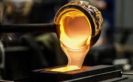
You can concentrate a large amount of molten glass on the end of a hollow tube, while rotating, while blowing air from the other end of the tube. The air entering the hollow tube, the action of gravity, and the tools used by the glass blower shapes the glass.

Pour the molten glass into a tank of tin bath, float on the surface of the relatively dense tin liquid, and slowly shape it under the action of pressurized nitrogen. The glass produced by this method is called float glass and has been used to produce flat glass since the 1950s.

Let the glass cool down. Do not place the glass in a place where it will be disturbed, dust, leaves, or water droplets. Exposing a cool substance to hot glass can cause the glass to burst.

The glass is strengthened by heat treatment. This process is called annealing and removes any stress points formed on the glass during the cooling process. Glass that has not been annealed is significantly weaker. Once this process is completed, the glass can then be coated, laminated, or other treatments to improve strength and durability.

The precise temperature of annealing varies, depending on the exact composition of the glass, as low as 399 degrees Celsius (750 degrees Fahrenheit) and as high as 538 degrees Celsius (1000 degrees Fahrenheit). [3] The cooling rate of glass is also different. Generally, the cooling rate of a larger piece of glass must be slower than that of a smaller glass. Investigate the correct annealing method before starting annealing.

Another related process is tempering. The polished shaped glass is heated to at least 600 degrees Celsius (1112 degrees Fahrenheit) in an oven and then rapidly cooled (quenched) with air under high pressure. Annealed glass will become fragments under a pressure of 6000 psi (421.94 kg/cm2), while tempered glass will become fragments under a pressure of no more than 10,000 psi (703.23 kg/cm2), usually about 24,000 psi (1687.76 kg/cm2).

To be continued in Part One…


Four Common Protein Interaction Techniques You Must Know

As the main bearer of life activities, the function of proteins has always been a star that has attracted much attention in scientific research activities. Proteins are usually not “fighting alone”, and the vast majority of functional proteins will interact with other proteins to regulate life processes together. So, how much do you know about the technology of protein interaction?I. Co-immunoprecipitationCo-immunoprecipitation (COIP) technique is based on the principle of specific immune binding between antigen and antibody. Specific antibody is added to cell lysis to precipitate antigen and antigen-bound protein. Immune complexes can verify the interaction between antigens and other proteins by Western blot, and can also detect the binding protein members of antigens by mass spectrometry.

Advantages and disadvantages of COIP technology:

Advantages

(1) The obtained interacting proteins are naturally interacting with bait proteins in cells, which are in line with the real physiological situation in vivo; (2) The experimental conditions are mild and artificial effects can be avoided; (3) The interacting protein complexes in the native state can be isolated.

Disadvantages:

(1) It is not applicable to the study of protein interaction with weak or instantaneous binding.

II. Pull Down

Pull-down techniques use solid-phase, labeled bait proteins or tag proteins (biotin-, PolyHis-, or GST-) to fish out proteins that interact with them from cell lysis. The interaction relationship between known proteins and phishing proteins or purified related proteins can be determined by pull-down technology, and the protein interaction relationship can be detected from in vitro transmission or translation systems. The protein interaction detected by COIP may be established by the third protein as a bridge, in contrast to the pull down technique, which can be used to detect direct interaction between proteins. However, pull down requires prokaryotic expression and purification of the bait protein, followed by incubation with the target protein solution, which cannot mimic the natural interaction environment in cells like COIP.

III. Bimolecular fluorescence complementationBimolecular fluorescence complementation technique refers to the incision of fluorescent protein polypeptide chains at some unconserved amino acids to form two non-fluorescent N- and C-terminal polypeptide fragments. These two fluorescent protein fragments were ligated to a pair of target proteins that could interact, respectively, and when these two fusion proteins were co-expressed intracellularly or mixed in vitro, due to the interaction of the target proteins, the two fragments of the fluorescent proteins were spatially close to each other and complementary to each other, and a complete active fluorescent protein molecule was reconstructed, thus producing fluorescence. Visualization is the greatest feature of BiFC technology.

IV. Yeast two-hybridYeast two-hybrid system is an important method widely used in protein interactomics. The principle is that when the target protein and the bait protein specifically bind, the bait protein binds to the promoter of the reporter gene and initiates the expression of the reporter gene in yeast cells, and if the expression product of the reporter gene is detected, it indicates that there is an interaction between the two, otherwise there is no interaction between the two. In practice, people have developed one-hybrid system, three-hybrid system and reverse hybridization system according to needs.

In addition, the methods to study protein interaction include phage display technology, plasmon resonance technology, antibody and protein array technology.

Collected by Profacgen

We have established in our lab a series of assays to help with your research, including high throughput interaction screening assays such as Yeast two-hybrid screening and phage display technology, interaction strength and kinetics assays such as Surface Plasmon Resonance (SPR).


3 Things You Must Know About Tetanus

Abstract: Today, tetanus, also known as lockjaw, is uncommon in the US, with only about 30 cases each year. However, due to its high fatality rate, it’s still necessary to know more about its causes, symptoms and prevention.

On sunny and warm days, everyone likes to be outdoors, but bumps, cuts and abrasions will inevitably occur, causing skin damage. If the wound is not treated in time, it may cause tetanus. So what is going on with tetanus? What is the use of tetanus vaccine?

·Causes and symptoms of tetanus

Tetanus is an acute infectious disease caused by tetanus bacteria invading human wounds. Once infected with this bacteria, the fatality rate is 20% to 40%. Tetanus infection usually has an incubation period of one week, and some individuals only develop symptoms within a day or months, even years. Nearly 90% of patients develop symptoms within two weeks after injury.

The main symptoms are damage to the central nervous system, manifesting continuous muscle contraction and other symptoms. The first is the masticatory muscles, then the face, neck, back, abdomen, limbs, and finally the diaphragm and intercostal muscles.Sound, light, vibration can all induce paroxysmal cramps. In severe cases, respiratory paralysis occurs which can cause death. The prevention of tetanus first emphasizes standardized wound treatment, while implementing artificial immunization.

·Situations where a tetanus vaccine is necessary

The simple criterion for tetanus vaccine is to answer these two questions: what’s the depth of the wound and what caused the wound?

When the wound is deep enough to require stitches, or when it suffers some serious trauma, such as nails, wood, thorns and other sharp materials, which cause bleeding, a vaccine is necessary because these kinds of deep and thin wounds are precisely hotbed for tetanus bacteria.

The wound, which has a small outer opening, or has necrotic tissue, clot filling or tight packing, ischemia, etc. in the wound, will become a hypoxic environment suitable for the growth and reproduction of this bacteria. If there is an infection ofaerobic bacteria at the same time, the latter will consume the residual oxygen in the wound, making tetanus more likely to occur.

It can be seen that tetanus infection usually does not occur in superficial wounds. For simple surface abrasions, after timely debridement, there is no need to inject the vaccine for prevention; but if the wound is deep and the pollution is serious, the possibility of tetanus will be greatly increased, and then the vaccine is necessary.

·Principle of tetanus vaccine

Tetanus prevention is clinically divided into active immunization and passive immunization. The so-called active immunization is to inject tetanus toxoid into the human body for 3 times, which can protect the patient for 2-3 years. Another situation is to inject patients with tetanus antitoxin, or immunoglobulin, which belong to passive immunity.

“Tetanus vaccine, also known as tetanus toxoid (TT), is a kind of immune horse serum, which is a heterogeneous protein to the human body. It is antigenic and may cause allergic reactions and other adverse reactions. Therefore, an allergy test should be done before medication.” introduced by a scientist at Creative Diagnostics, a biotech company experting in various bacterial antigens.

If the test result is positive, tetanus antitoxin should be injected, through 4 to 5 injections in small doses. For those who have received a test for more than 1 week, the skin test must be repeated if they are used again.

Tetanus immunoglobulin generally causes no allergic reaction, so there is no need to test before injection. It is just a kind of exogenous antibody, which can be maintained in the body for 2 to 3 weeks. It should be used in time when suspicious symptoms have appeared, but the preventive effect of tetanus is limited.

·Two common misunderstandings of vaccination

The first misunderstanding is that the injection is effective only within 24 hours after trauma. The incubation period after infection is mostly 1 to 2 weeks. According to its pathogenesis, application of tetanus antitoxin within 24 hours or even later after injury can play a preventive effect. It is suggested to inject the vaccine as soon as possible, but as long as the symptoms do not occur, the vaccination within 2 weeks should be regarded as a preventive effect.

The second misunderstanding is that people do not have to receive another injection after they once have received one. As it is said in the previous chapter, the vaccine will not give our bodies lasting immunity, thus following the doctor’s advice is the best way to protect ourselves.


How to enhance the therapeutic efficiency of anti-PD-L1 cancer immunotherapy?

For most people, nothing is more terrible than cancer. Therapies such as chemotherapy and radiotherapy began to be used in the 1940s and late 1800s, respectively, while immunotherapy has only been used in recent years as a viable and successful treatment. Indeed, evading the host immune system is a basic feature of tumorigenesis, and it may be very important to elucidate how cancer cells do this, and how to promote the patient’s own immune system to eliminate cancer cells, which may also be the basis for new immunotherapies.

Recently, in a study published in Nature Cell Biology, scientists from Tokyo Medical and Dental University and other institutions identified a special regulatory mechanism, that is, how PD-L1 immune checkpoint proteins affect the efficiency of anti-PD-L1 immunotherapy. Now we know that immunotherapy against immune checkpoint inhibitors can successfully treat certain cancer types, but only a small proportion of patients can have durable treatment outcomes, the researchers said.PD-L1 expression is strictly controlled, and patients with tumors carrying increased PD-L1 expression levels respond well to PD-L1 arrest. However, it remains unclear why increasing PD-L1 expression leads to increased sensitivity to PD-L1 arrest. Subsequently, the researchers analyzed one type of PD-L1 modification, acetylation modification, and found that removing this modification may promote PD-L1 to enter the nucleus and interact with DNA to regulate the immune response of the host body.

Using a variety of advanced molecular, biochemical, and bioinformatics analysis tools, the researchers analyzed the acetylation modification, localization characteristics, function, and interaction mechanism of PD-L1 and found that plasma membrane-localized PD-L1 could be transferred into the nucleus by interacting with transport pathway components.Specifically,by introducing a series of mutations in PD-L1 and expressing different acetyltransferases, the researchers found that PD-L1 could be acetylated by a special residue called Lys262 in the cytoplasm, using similar methods and protein removal mediated by short interfering RNAs, and the researchers also found that histone acetylase (HDAC) could specifically interact with PD-L1 and deacetylate it.Protein modification including acetylation modification can affect protein stability, dimerization and localization. However, when investigators reduce the expression of HDAC2 protein in cells, they increase the level of acetylation, but they do not observe changes in protein stability or dimerization. The relevant study results show that acetylation modification and deacetylation of PD-L1 at specific residue sites play a key role in the nuclear transport process; in the nucleus, PD-L1 can regulate the expression of inflammatory and immune response-related genes, suggesting that PD-L1 can function to regulate the local tumor immune environment and thus control its sensitivity to immune checkpoint blockade therapy.Finally, the researchers say, considering the health and economic burden of cancer populations worldwide, researchers need to conduct more in-depth studies to find new anti-cancer therapies, and the results of this study suggest that targeting the translocation of PD-L1 may help enhance the therapeutic efficiency of PD-1/PD-L1 block-based immunotherapy.

Collected by Creative BioMart

Creative BioMart presents immune checkpoint proteins developed as recombinant proteins. These products cover multiple species and are available in various expression hosts. Bio-activity of all immune checkpoint proteins has been assayed and confirmed, making them essential materials for mechanistic studies and development of new therapeutics.


An Overview of Researches on Targeted Protein Degradation (I)

Interference with specific protein expression is an important way to study protein function, and it is an important measure to treat related diseases caused by abnormal protein expression. From the earliest chemical inhibitors that work by blocking the active site of the target protein to the RNAi technology and CRISPR-Cas9 technology based on the level of DNA and RNA, they all play the function of knocking down the target protein to a certain extent. However, with the deepening of research, the limitations of the technology have been exposed: chemical inhibitors can only exert effects when they are combined with the active site of the target protein under the action of high concentrations of compounds, and have high cytotoxicity. Although RNAi technology and CRISPR-Cas9 technology solve the shortcomings of chemical inhibitors to a certain extent, they are prone to off-target effects. As an indirect method, it takes a long time to interfere with the expression of specific proteins, which causes cells to have enough time to activate the compensation mechanism and restore the original for some protein levels, so it is impossible to determine whether the decrease in target protein expression is related to early defects at the gene level.

Targeted protein degradation technology (PROTAC) is a new technology developed in recent years that interferes with protein function and maintains the homeostasis of intracellular proteins. Targeted protein degradation technology is one of the most direct and effective ways to regulate protein expression. It can specifically identify the target protein and use the inherent protein degradation pathway in the cell to directly degrade the target protein at the protein level, thereby improving the efficiency of knocking out the target protein. And the omission of the intermediate effects produced during the long-term regulation process provides a new method for studying the function of specific proteins and treating diseases caused by abnormal protein expression, so it has gradually attracted widespread attention.

The principles of targeted protein degradation technology

The protein homeostasis in the cell mainly depends on the coordinated operation and maintenance of the inherent protein degradation pathways in the cell, that is, the molecular chaperone protein system and two proteolytic systems, among which the autophagy-lysosome pathway  and the ubiquitin-proteasome pathway (UPP) play an important biological function in protein degradation.

1.1 Autophagy-lysosomal pathway

The autophagy-lysosome pathway is a non-specific protein degradation pathway, which is mainly related to the degradation of surface membrane proteins, endocytosed extracellular proteins, intact organelles and protein multimers, and does not play a major role in regulating intracellular protein degradation. Lysosomes are important organelles in eukaryotic cells and are an important component of the inner membrane system. They contain a variety of acid hydrolases that can decompose proteins, nucleic acids, polysaccharides and lipids. Its main mechanism of action is: when cells are stimulated by conditions such as starvation, pressure, high temperature, hypoxia, organ damage, protein mutations, or microbial infections, autophagy will be triggered, and large molecules such as proteins will be affected by the acidic environment of the lysosome. Corresponding enzymes are degraded and then transported to the cytosol metabolic pool through the carrier protein of the lysosomal membrane.

1.2 Ubiquitin-proteasome pathway

The ubiquitin-proteasome pathway is the most important protein degradation pathway in the cell. It is highly specific and selective. It is a key regulator of the stability of the intracellular environment. It is responsible for degrading 80% of normal or abnormal proteins in cells and in the cell cycle. And it plays an important role in cell regulation, signal transduction, nucleic acid code translation and other life processes, and these are the main ways to participate in targeted protein degradation technology in cells. The ubiquitin-proteasome pathway mainly includes: ubiquitin (Ub), ubiquitin-activating enzyme (E1), ubiquitin-conjugating enzyme (E2), ubiquitin ligase E3, 26S proteasomes, deubiquitinating enzymes (DUBs), etc. The main mechanism of action of the ubiquitin-proteasome pathway (Figure 1): E1 uses the energy released by ATP hydrolysis to activate a single free ubiquitin through the formation of a thioester bond between the cysteine residue in its active center and the C-terminus of ubiquitin, and then present ubiquitin to E2; E3 has the ability to specifically recognize the target protein, which mediates the transfer of ubiquitin from E2 to the target protein and covalently binds to the target protein by recruiting substrate proteins and E2, and the proteasome catalyzes ubiquitination. The action process of the ubiquitin-proteasome pathway is a reversible process, which maintains the balance through E1, E2, E3 and DUBs. Mono-ubiquitination cannot mediate the degradation of the target protein, but only plays a certain degree of regulation. Only when 4 or more activated ubiquitin molecules are connected to the target protein to form a polyubiquitin chain, can the substrate be transported to the proteasome for degradation, and the polyubiquitin chain is decomposed into individual ubiquitin molecules under the action of the ubiquitin recycling enzyme, and then recycled.

Figure.1 The main reaction mechanism of ubiquitin-proteasome pathway

2 2 Basic structure and mechanism of PROTAC

As a bifunctional molecule, PROTAC includes three parts: one end is a ligand that targets the target protein; the other end is a structure that can recruit protein degradation systems (such as E3 ligase); the middle part is connected by a suitable linking chain, which is essentially one kind of splicing. The mechanism of PROTAC-induced protein degradation is shown in Figure 2. After PROTAC enters the cell, the protein of interest (POI) ligand in its structure specifically binds to the target protein, and the E3 ligase ligand binds to E3 ligase to form a POI-PROTAC-E3 ligase ternary complex. E3 ligase mediates the ubiquitination of the target protein by the ubiquitin-conjugating enzyme E2. After the ternary complex is dissociated, the target protein labeled with ubiquitin will be transported to the proteasome for degradation, thereby selectively reducing the level of the target protein. In this process, the target protein ligand does not need to occupy the binding site for a long time, and only needs to form a ternary complex briefly to complete the ubiquitination of the target protein. Protein with ubiquitination tag can be recognized by the proteasome and be degraded. Moreover, PROTAC plays an important role in multiple cycles within the cell.

Figure 2. Direct recruitment of an E3 ligase by using the PROTAC

3 Ubiquitin E3 ligase-based protein degradation targeted chimera

  1. 1 Peptide PROTAC

Due to the lack of small molecule ligands for E3 ubiquitin ligase, the first generation of PROTAC based on peptide motifs is used as the ligand for E3 ubiquitin ligase. Although this type of molecule provides a proof of concept for PROTAC, these peptides sequences lack cell permeability, thus limiting their utility as chemical probes. Studies have confirmed that the target proteins that peptide PROTAC can successfully degrade include androgen receptor (AR), methionyl aminopeptidase 2 (MetAP2) and protein kinases (AKT, PI3K), etc. These studies confirmed that targeting E3 ligase to regulate the half-life of proteins is a potential strategy for drug development. However, these peptides PROTAC have poor membrane absorption and low protein degradation activity, and their degradation activity is still at the micromolar level.

To be continued in Part II…


Liposomes: An Excellent Partner of Antiprotozoal Drug

Parasitic diseases such as malaria, leishmaniasis, and trypanosomiasis are still problems that threaten human health. About 30% of the world’s population is infected with parasitic diseases. Most parasitic diseases do not trigger an immune response and thus no suitable vaccine can be found. Therefore, the treatment of parasitic diseases depends more on drug treatment. The disadvantage of traditional antiprotozoal drugs is that they cannot target cells. Repeated administration of large doses leads to multiple drug resistance and drug toxicity.

Taking into account the infection characteristics of protozoal diseases, the drug delivery system of antiprotozoal drugs should meet the following requirements:

1. Able to achieve oral administration;

2. Intracellular targeting, high drug loading rate, low toxicity, and low immunogenicity with the ability to protect the drug from being degraded before it enters the cell;

3. Improve the pharmacodynamic parameters of antiprotozoal drugs and reduce the administration cycle;

4. Transport the combination of antiprotozoal drugs and other substances flexibly;

5. The cost-benefit ratio;

6. Targeting Delivery of antiprotozoal drugs to achieve maximum efficacy and minimum adverse reactions.

Figure 1. Transmission electron Microscopy. Notes: (A) Plain liposome; (B) liposomes loaded with vancomycin; (C) Tat-functionalized liposome loaded with vancomycin.

· Liposome for Anti-malaria Drug

In the 1980s, scientists have prepared liposome formulations of antimalarial drugs such as quinine, chloroquine, primaquine, artemether, and artesunate. The pH gradient method is a good way to improve the encapsulation efficiency of antimalarials, especially for quinolones. The chloroquine PEGylated long-circulating liposome prepared by the pH gradient method released 30% of the drug within 6 hours at a pH of 7.4, and over 90% of the drug was released at a pH of 5.5. The artesunate liposomes prepared by using a buffer solution of pH 5 as the aqueous phase can reach almost 100% in the encapsulation efficiency, which can improve the stability of artesunate.

The relative oral bioavailability of artemether liposomes can reach 97.9%, while the artemether suspension is only 31.8%. Primaquine anionic liposomes prolong the plasma half-life of the drug, and the clearance rate is reduced by 8 times. About 50% of the injected drug is distributed in the liver, which is twice the free drug, and the spleen intake is increased by 3 times. The distribution in the lung, kidney, heart and brain is significantly reduced, thus decreasing the systemic toxicity of the drug. Scientists have prepared liposomes modified with monoclonal antibodies that specifically bind to erythrocytes infected with Plasmodium, and encapsulated with chloroquine, and then were injected into mice infected with Plasmodium. After administration for 4 days and 6 days, the cure rates were 75% and 90%, respectively, which means the liposome is not only effective against drug-sensitive plasmodium, but also effective against drug-resistant plasmodium.

· Liposome for Anti-leishmania Drug

Because Leishmania reproduces in macrophages, and macrophages are an important place for liposome elimination, liposomes are the most effective treatment for leishmaniasis.

The biological activity of sodium antimony gluconate liposomes against experimental leishmaniasis in hamsters is 700 times that of free drugs. The decomposition of Leishmania in Kupffer cells is clearly observed under the electron microscope, which fully reflects the broad prospects of liposome drug delivery system in the treatment of leishmaniasis. Sodium antimony gluconate liposomes are also effective against skin leishmaniasis.

In terms of macrophage drug uptake and targeting liver and spleen, cationic liposomes have more advantages than anionic and neutral liposomes. Cationic liposomes composed of phosphatidylcholine and stearylamine encapsulate sodium antimony gluconate, which can significantly reduce the amount of Leishmania in the liver. The mechanism may be the electrostatic effect of cationic liposomes and protozoan plasma membrane, which destroys the organelle structure and inhibit the oxygen consumption of the protozoa.

The macrophage membrane has receptors that recognize mannosyl, galactosyl, fucose residues and glucose residues of glycosides. Mannose modified urea antimony amine liposomes can more effectively deliver drugs to macrophages, and also more effectively reduce the amount of parasites in the spleen and drug toxicity. Glycoprotein-modified harmycin liposomes have a clearance rate of intracellular amastigo-protozoa that is twice that of ordinary liposomes and 10 times that of free drugs. Glycoprotein modified liposomes can completely eliminate the protozoa in the spleen, while ordinary liposomes can only partially eliminate them. Macrophage activating peptides, such as phagocytic peptides, are used to improve the targeting of anti-leishman drugs due to their preferential binding to the mononuclear macrophage system. There are Fc receptors on the surface of macrophages, and liposomes can be combined with some Fc antibodies such as immunoglobulins (Ig) to form immunoliposomes, which are used to treat Leieman’s disease. When sodium antimony gluconate is administered, the antibody has a synergistic effect against Leishman. Therefore, modified liposomes with this antibody can not only improve the targeting of macrophages, but also have anti-Leishman synergistic effects.

· Liposome for Anti-trypanosomiasis drug

Trypanosomal pathogens are distributed in general cells, not in the mononuclear macrophage system, so liposomes are rarely used in the treatment of trypanosomiasis. But blank cationic liposomes have the potential to resist trypanosomiasis. Cationic liposomes composed of phosphatidylcholine and stearylamine can kill trypanosomes within 30 minutes at a very low lipid concentration (100μmol/L) and have no toxicity to red blood cells. The mechanism may be to destroy plasma membrane stability of trypanosomes, while intervening in the dimensional mechanism of trypanosomal plasma membrane.

The second-generation nitroimidazole drug, etanterol, is prepared into pH-sensitive liposomes, which can avoid the endocytosis of lysosomes, significantly improve the cytoplasmic targeting of the drug, and clear 72% of trypanosomes in infected macrophages within 2 hours, while free drugs and ordinary liposomes do not have any antitrypanosomal activity. Arsenic compounds, such as melarsoprol, can be used to treat trypanosomal infections. Arsenic liposomes have significant anti-trypanosomal activity at lower concentrations, and their porous structure is very important for their anti-trypanosomal effects.


A Research about Induced Pluripotent Stem Cells and Genome Editing Techniques for Cardiovascular Disease

Abstract

Cardiovascular disease is the manifestation of systemic vascular disease or systemic vascular disease in the heart and brain. It is the top one cause of death worldwide. Cardiovascular diseases can cause hemiplegia and even death, and bring serious economic burdens to patients and their families. Therefore, cardiovascular disease research is also a global concern.

The prevention of cardiovascular and cerebrovascular diseases includes primary prevention and secondary prevention. Primary prevention refers to prevention before the onset, which is, preventing disease from occurring; secondary prevention is to reduce the risk of recurrence and reduce the disability rate, that is, prevent recurrence after illness. A recent study has studied the treatment of cardiovascular diseases through the direction of genetics and genomics.

The study was published in “Cell Reports” on September 9 with the title: “iPSC Modeling of RBM20-Deficient DCM Identifies Upregulation of RBM20 as a Therapeutic Strategy”. The researchers found that: for familial dilated cardiomyopathy (DCM) for patients, using drugs such as ATRA to increase the level of RBM20 in heart cells is a promising treatment option.

Dilated cardiomyopathy (DCM) can cause heart failure. DCM is characterized by increased blood volume in the left ventricle of the heart (the main pump chamber), and the stretched myocardium cannot pump blood. This can lead to arrhythmia, heart valve problems, and eventually heart failure. DCM is not only the main cause of heart failure, but also the most common cause of heart transplantation. Heart transplantation is only considered when other treatment options and lifestyle changes have failed in the late stages of heart failure.

Despite continuous efforts to improve the survival rate of heart transplant patients, the 10-year survival rate is still only 50%. Therefore, if there are better treatment options before heart transplantation, the health of DCM patients will be significantly improved.

Many DCMs are caused by genetic changes (mutations) in human DNA. Although there is a detailed understanding of mutations, there is no therapy for mutations. Modeling the ability of induced pluripotent stem cells (iPSC-CMs) to produce cardiomyocytes will enable people to better understand hereditary heart disease and seek treatments for it.

A team of researchers led by Dr. Francesca Briganti, Dr. Han Sun, senior authors Dr. Mark Mercola, Dr. Lars Steinmetz, and Dr. Ioannis Karakikes of Stanford University, combined iPSC-CM and genome editing to identify new mutations that cause DCM, and found DCM is better treatment.

Researchers conduct studies on families who have inherited DCM to understand its cause. As a genetic disease, genome information or complete DNA and genetic information can provide important information about mutations that cause the disease. Each gene contains a specific protein required to make cells, and the gene is passed from the parent to the child, including protein changes that cause diseases such as inherited DCM.

Family hereditary dilated cardiomyopathy has specific changes in RBM20

In this case, the research team compares the fragments of the genome that make up the protein (exome). By studying different families, they were able to find the exome differences between people who died due to a diagnosis of DCM, were diagnosed with DCM, and those who were not affected by DCM. By comparison, they can find a single mutation (P633L) in the protein RBM20, which causes disease. Although the mutation is unknown, it is known that RBM20 changes can cause the inheritance of DCM. The researchers also found that when one copy of a gene is changed (mutated) and the other copy is normal, the normal RBM20 function is lost, which is called haploinsufficiency.

iPSC, especially the iPSC-CM platform, is a powerful tool for studying specific cardiovascular diseases (such as genetic DCM). Researchers can use genome editing (a process that makes specific changes to the DNA of a cell) to introduce the RBM20 mutation (P633L) into iPSC-CM. This gives them an opportunity to understand how mutations cause DCM.

Importantly, the iPSC-CM platform also allows researchers to test the use of all-trans retinoic acid (ATRA) as a potential treatment for DCM by adjusting RBM20. In the body, ATRA is made of vitamin A, which helps cells grow and develop. Interestingly, ATRA treatment can increase the level of RBM20. More RBM20 can restore function to normal levels, thereby treating genetic DCM caused by mutations (under-dose). Here, the researchers used iPSC-CM with a specific RBM20 mutation (P633L) to prove that ATRA can partially repair defects in the changed cells. It is crucial that researchers can prove that for patients with familial DCM, using drugs such as ATRA to increase the level of RBM20 in heart cells is a promising treatment option.

About genome editing

Genome editing involves the insertion, deletion, modification or replacement of DNA in the genome with the help of synthetic nucleases, the “molecular scissors”. Targeting genome alteration of stem cells in disease models is a prerequisite for utilizing the full potential of stem cells. Genome edited human iPSCs can be employed to study gene function, drug and chemical screens. Many genome-editing techniques have been launched to improve the efficiency and speed of the development of stem cells for human disease models.

Genome edited human pluripotent stem cells hold great promise for gene therapy approaches. The development of efficient technologies for stem cell genome editing has attracted massive attention. There are various technologies have been developed to provide high-quality gene editing services:

CRISPR

TALEN

Recombinant DNA

* ZFN technology