

Il mondo non è omogeneo. È ricco di culture e comunità ricche, ognuna delle quali è unica in qualche modo nella sua ideologia, valori e altro ancora. Anche se il mondo è diventato più interconnesso con l’avvento del World Wide Web e una maggiore facilità di viaggio, il confronto prevalente tra “Oriente” e “Occidente”, Asia ed Europa (o ora, Europa e America), ha continuato a essere oggetto di studio per centinaia di anni (non importa quanto il confronto tra i due sia diventato obsoleto e problematico). Questo continuo confronto sembra essere bloccato ai tempi di Marco Polo, quando potevamo mandare qualcuno in Oriente – “in Asia!” – riportare notizie, merci e altre “scoperte”. Ora, le nostre scoperte prendono la forma di articoli scientifici. Qui esaminerò uno di questi documenti, uno studio del 2012 di Murata, Moser e Kitayama.
Lo studio:
Abbiamo sempre saputo che la cultura modella chi siamo, ma ora, con metodi neuroscientifici, possiamo esaminare quanto la cultura può modellare il nostro cervello . Il documento del 2012 “La cultura modella le risposte elettrocorticali durante la soppressione delle emozioni” di Murata, Moser e Kitayama mirava a esaminare un aspetto dell’influenza della cultura sul modo in cui funziona il nostro cervello, in particolare su come elaboriamo e sperimentiamo le emozioni.
Contesto: l’emozione fa parte dell’esperienza umana, ma alcune situazioni possono richiedere una regolazione delle emozioni in modo che non prevalga sui processi di pensiero più razionali. Questo si chiama controllo emotivo, cioè avere il controllo delle proprie emozioni. Esistono diversi modi per farlo: consapevolezza, rivalutazione, ecc. Murata, Moser e Kitayama hanno esaminato la soppressione emotiva, ovvero il processo faticoso di sopprimere, o lavorare per diminuire e persino eliminare, le emozioni. Sebbene le persone di tutte le culture possano riconoscere gli affetti negativi e positivi (cioè le emozioni tristi e felici) in base ai volti delle persone, culture diverse apprezzano l’espressione delle emozioni in modo diverso. Le culture asiatiche, ad esempio, apprezzano il controllo delle emozioni, mentre le culture europee americane tendono a valutare maggiormente l’espressione delle emozioni. Murata, Moser e Kitayama ha cercato di valutare se queste diverse valutazioni del controllo emotivo si riflettessero nelle risposte elettrocorticali delle persone durante un esperimento. In altre parole, volevano scoprire se questa preferenza fosse evidente nell’attività neurale. I ricercatori hanno ipotizzato che le persone che crescono nelle culture asiatiche siano “educate culturalmente” a sopprimere meglio e più frequentemente le proprie emozioni, rispetto agli americani europei che hanno “meno probabilità” di avere un simile addestramento alla soppressione emotiva.
Obiettivi dello studio: i ricercatori hanno utilizzato un potenziale parietale tardivo positivo (LPP) del potenziale correlato all’evento (ERP) (una tecnica di misurazione del cervello) per esaminare le differenze nell’attività neurale nel prestare attenzione (cioè prestare attenzione) o sopprimere le emozioni tra persone di culture diverse, in particolare culture asiatiche e culture americane europee.
Come ha funzionato lo studio: ai partecipanti è stata mostrata una serie di immagini che suscitavano un effetto negativo (es. immagini di mutilazione, altre immagini spiacevoli) o erano neutre (es. immagini di volti o scene neutre). I partecipanti sono stati istruiti a prestare attenzione (prestare attenzione) alle loro emozioni e reagire normalmente o a sopprimere le loro emozioni e diminuire le loro risposte emotive sia internamente che esternamente (cioè mediante l’espressione facciale, ecc.)
Risultati e conclusioni dello studio:
-Entrambe le culture hanno mostrato un’attività neurale simile dopo la presentazione delle immagini, indicando che tutti avevano una reazione emotiva iniziale simile agli stimoli.
-Quando sono stati istruiti a sopprimere le loro emozioni, i soggetti asiatici hanno mostrato una significativa diminuzione dell’attività neurale associata alla loro emozione, con conseguente scomparsa totale di quella positività neurale iniziale in pochi secondi. Ciò suggerisce che i soggetti asiatici sono abbastanza abili nel sopprimere le proprie emozioni.
-I soggetti europei americani hanno dimostrato di essere meno abili nel sopprimere le proprie emozioni. È interessante notare che c’è stato anche un aumento sostenuto della positività frontale (che potrebbe essere vista come “più” emozione) quando stavano effettivamente cercando di sopprimere/regolare le loro emozioni.
-I ricercatori hanno concluso che la loro ipotesi di “addestramento culturale” con conseguente soppressione emotiva più abile nei soggetti asiatici era corretta.
La mia opinione:
Vorrei che gli autori dello studio discutessero maggiormente della loro idea di “formazione culturale”. Non siamo tutti culturalmente formati? Ho discusso di questo studio con diversi amici che sono cresciuti in varie parti dell’Asia o sono stati cresciuti da genitori asiatici, e hanno discusso delle differenze nell’istruzione (cioè essere scoraggiati dal parlare e fare domande) così come le differenze nell’educazione (come non sentire mai i loro genitori dicono “ti amo”). Mi chiedo cosa significhi “formazione culturale” qui, ma sono anche molto preoccupato per l’idea che gli asiatici sembrano ricevere una formazione culturale mentre gli americani europei no, il che normalizza l’esperienza degli americani europei come la normale linea di base. Sentire le emozioni è normale – e umano – così come il bisogno di controllarle.
Questo studio è pesante e scorretto nella generalizzazione delle culture. I partecipanti provenivano da una varietà di paesi dell’Asia orientale, tra cui Cina, Giappone e Singapore. Altri paesi in Asia, come India e Turchia, non sono stati inclusi, sebbene i risultati possano essere interpretati come generali anche a loro. Le differenze culturali tra i diversi paesi, e in effetti, all’interno dei paesi, sono vaste: considerarli abbastanza omogenei non riesce a catturare ciascuna delle loro culture, così come i diversi tratti della personalità di ciascuno degli individui studiati. Sfido anche l’assunto che le culture europee americane valorizzino di più l’espressività emotiva – nella mia esperienza, tendiamo ad ammirare coloro che possono controllare le proprie emozioni, lamentandosi nel frattempo che non possiamo fare lo stesso.
Le culture non esistono nei binari e potrebbe essere pericoloso continuare a perpetuare questa visione, specialmente quando il mezzo più oggettivo per la scoperta e l’esame, la scienza, continua a utilizzare questa retorica. Ciò è evidente nel testo stesso tra virgolette come gli asiatici che usano “soppressione automatica e senza sforzo” nonostante i loro resoconti utilizzino lo stesso sforzo delle controparti del loro studio e la paura che gli asiatici possano essere “indotti a provare emozioni”. La questione che dovrebbe essere in gioco qui è come le diverse ideologie che vengono insegnate alle persone nel corso della loro vita e le abilità che apprendono di conseguenza influenzano la loro attività neurale, non come affrontare al meglio le differenze culturali. Vorrei non sentirmi in dovere di parlarne, ma anche se questo studio ha molti contributi interessanti e importanti-continua
Memorie.
BIBLIOGRAFIA
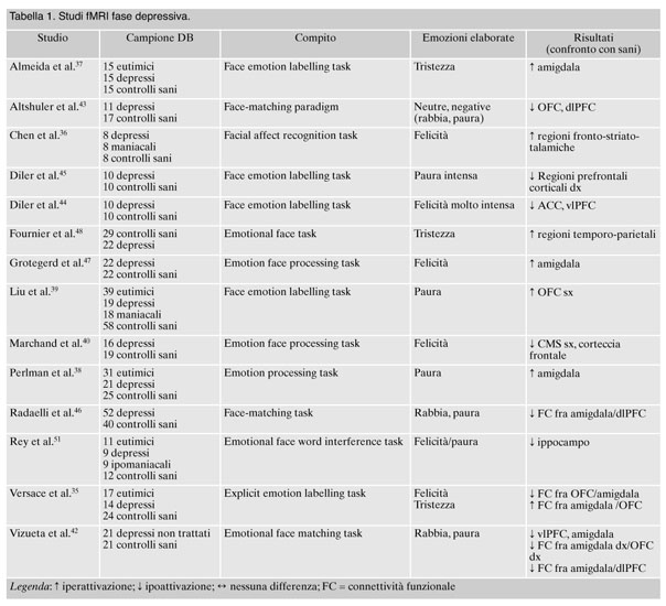
1. Cusi AM, Nazarov A, Holshausen K, Macqueen GM, McKinnon MC. Systematic review of the neural basis of social cognition in patients with mood disorders. J Psychiatry Neurosci 2012; 37: 154-69.
2. Couture SM, Penn DL, Roberts DL. The functional significance of social cognition in schizophrenia: a review. Schizophr Bull 2006; 32 (Suppl 1): S44-63.
3. Blundo C. Neuroscienze cliniche del comportamento. Basi neurobiologiche e neuropsicologiche. Psicopatologia funzionale e neuropsichiatria. Milano: Elsevier, 2011.
4. Batson CD. Two forms of perspective taking: imagining how another feels and imagining how you would feel. In: Markman KD, Klein WMP, Suhr JA (eds). Handbook of Imagination and Mental Stimulation. New York (NY): Psychology Press, 2009.
5. Davis MH. Measuring individual differences in empathy: evidence for a multidimensional approach. J Pers Soc Psychol 1983; 44: 113-26.
6. Leiberg S, Anders S. The multiple facets of empathy: a survey of theory and evidence. Prog Brain Res 2006; 156: 419-40.
7. Singer T. The neuronal basis and ontogeny of empathy and mind reading: review of literature and implications for future research. Neurosci Biobehav Rev 2006; 30: 855-63.
8. Adolphs R. The social brain: neural basis of social knowledge. Annu Rev Psychol 2009; 60: 693-716.
9. Ongür D, Price JL. The organization of networks within the orbital and medial prefrontal cortex of rats, monkeys and humans. Cereb Cortex 2000; 10: 206-19.
10. Rankin KP, Kramer JH, Miller BL. Patterns of cognitive and emotional empathy in frontotemporal lobar degeneration. Cogn Behav Neurol 2005; 18: 28-36.
11. Stone VE, Baron-Cohen S, Knight RT. Frontal lobe contributions to theory of mind. J Cogn Neurosci 1998; 10: 640-56.
12. Anderson AK, Phelps EA. Lesions of the human amygdala impair enhanced perception of emotionally salient events. Nature 2001; 411: 305-9.
13. Morris JS, Ohman A, Dolan RJ. Conscious and unconscious emotional learning in the human amygdala. Nature 1998; 393: 467-70.
14. Whalen PJ, Rauch SL, Etcoff NL, McInerney SC, Lee MB, Jenike MA. Masked presentations of emotional facial expressions modulate amygdala activity without explicit knowledge. J Neurosci 1998; 18: 411-8.
15. Adolphs R. The neurobiology of social cognition. Curr Opin Neurobiol 2001; 11: 231-9.
16. Altshuler LL, Ventura J, van Gorp WG, Green MF, Theberge DC, Mintz J. Neurocognitive function in clinically stable men with bipolar I disorder or schizophrenia and normal control subjects. Biol Psychiatry 2004; 56: 560-9.
17. Decety J, Lamm C. The role of the right temporoparietal junction in social interaction: how low-level computational processes contribute to meta-cognition. Neuroscientist 2007; 13: 580-93.
18. Olson IR, Plotzker A, Ezzyat Y. The Enigmatic temporal pole: a review of findings on social and emotional processing. Brain 2007; 130 (Pt 7): 1718-31.
19. Fusar-Poli P, Placentino A, Carletti F, et al. Functional atlas of emotional faces processing: a voxel-based meta-analysis of 105 functional magnetic resonance imaging studies. J Psichiatry Neurosci 2009; 34: 418-32.
20. Atkinson AP, Adolphs R. The neuropsychology of face perception: beyond simple dissociations and functional selectivity. Philos Trans R Soc Lond B Biol Sci 2011; 366: 1726-38.
21. Phillips ML, Drevets WC, Rauch SL, Lane R. Neurobiology of emotion perception I: the neural basis of normal emotion perception. Biol Psychiatry 2003; 54: 504-14.
22. Ochsner KN, Gross JJ. Thinking makes it so: a social cognitive neuroscience approach to emotion regulation. In: Baumeister RF, Vohs KD (eds). Handbook of self-regulation: research, theory, and applications. New York (NY): Guilford Press, 2004.
23. Phan KL, Fitzgerald DA, Nathan PJ, Moore GJ, Uhde TW, Tancer ME. Neural substrates for voluntary suppression of negative affect: a functional magnetic resonance imaging study. Biol Psychiatry 2005; 57: 210-9.
24. Ochsner KN, Ray RD, Cooper JC, et al. For better or for worse: neural systems supporting the cognitive down- and up-regulation of negative emotion. Neuroimage 2004; 23: 483-99.
25. Gusnard DA, Raichle ME. Searching for a baseline: functional imaging and the resting human brain. Nat Rev Neurosci 2001; 2: 685-94.
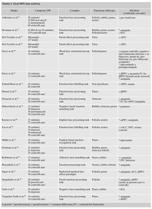
26. Tesli M, Kauppi K, Bettella F, et al. Altered brain activation during emotional face processing in relation to both diagnosis and polygenic risk of bipolar disorder. PLoS One 2015; 10: e0134202.
27. Brotman MA, Tseng WL, Olsavsky AK, et al. Fronto-limbic-striatal dysfunction in pediatric and adult patients with bipolar disorder: impact of face emotion and attentional demands. Psychol Med 2014; 44: 1639-51.
28. Horacek J, Mikolas P, Tintera J, et al. Sad mood induction has an opposite effect on amygdala response to emotional stimuli in euthymic patients with bipolar disorder and healthy controls. J Psychiatry Neurosci 2015; 40: 134-42.
29. Hulvershorn LA, Karne H, Gunn AD, et al. Neural activation during facial emotion processing in unmedicated bipolar depression, euthymia, and mania. Biol Psychiatry 2012; 71: 603-10.
30. Surguladze SA, Marshall N, Schulze K, et al. Exaggerated neural response to emotional faces in patients with bipolar disorder and their first-degree relatives. Neuroimage 2010; 53: 58-64.
31. Del Vecchio G, Fossati P, Boyer P, et al. Common and distinct neural correlates of emotional processing in Bipolar Disorder and Major Depressive Disorder: a voxel-based meta-analysis of functional magnetic resonance imaging studies. Eur Neuropsychopharmacol 2012; 22: 100-13.
32. Del Vecchio G, Sugranyes G, Frangou S. Evidence of diagnostic specificity in the neural correlates of facial affect processing in bipolar disorder and schizophrenia: a meta-analysis of functional imaging studies. Psychol Med 2013; 43: 553-69.
33. Foland-Ross LC, Bookheimer SY, Lieberman MD, et al. Normal amygdala activation but deficient ventrolateral prefrontal activation in adults with bipolar disorder during euthymia. Neuroimage 2012; 59: 738-44.
34. Favre P, Baciu M, Pichat C, et al. Modulation of fronto-limbic activity by the psychoeducation in euthymic bipolar patients. A functional MRI study. Psychiatry Res 2013; 214: 285-95.
35. Versace A, Thompson WK, Zhou D, et al. Abnormal left and right amygdala-orbitofrontal cortical functional connectivity to emotional faces: state versus trait vulnerability markers of depression in bipolar disorder. Biol Psychiatry 2010; 67: 422-31.
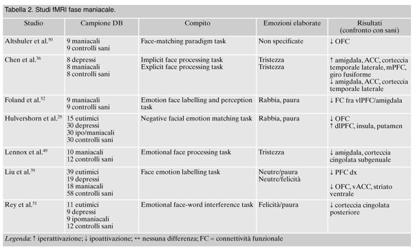
36. Chen CH, Lennox B, Jacob R, et al. Explicit and implicit facial affect recognition in manic and depressed states of bipolar disorder: a functional magnetic resonance imaging study. Biol Psychiatry 2006; 59: 31-9.
37. Almeida JR, Versace A, Hassel S, Kupfer DJ, Phillips ML. Elevated amygdala activity to sad facial expressions: a state marker of bipolar but not unipolar depression. Biol Psychiatry 2010; 67: 414-21.
38. Perlman SB, Almeida JRC, Kronhaus DM, et al. Amygdala activity and prefrontal cortex-amygdala effective connectivity to emerging emotional faces distinguish remitted and depressed mood states in bipolar disorder. Bipolar Disord 2012; 14: 162-74.
39. Liu J, Blond BN, van Dyck LI, Spencer L, Wang F, Blumberg HP. Trait and state corticostriatal dysfunction in bipolar disorder during emotional face processing. Bipolar Disord 2012; 14: 432-41.
40. Marchand WR, Lee JN, Garn C, et al. Aberrant emotional processing in posterior cortical midline structures in bipolar II depression. Prog Neuropsychopharmacol Biol Psychiatry 2011; 35: 1729-37.
41. Sagar KA, Dahlgren MK, Gönenç A, Gruber SA. Altered affective processing in bipolar disorder: an fMRI study. J Affect Disord 2013; 150: 1192-6.
42. Vizueta N, Rudie JD, Townsend JD, et al. Regional fMRI hypoactivation and altered functional connectivity during emotion processing in nonmedicated depressed patients with Bipolar II. Disorder Am J Psychiatry 2012; 169: 831-40.
43. Altshuler L, Bookheimer S, Townsend J, et al. Regional brain changes in bipolar I depression: a functional magnetic resonance imaging study. Bipolar Disord 2008; 10: 708-17.
44. Diler RS, Ladouceur CD, Segreti AM, et al. Neural correlates of treatment response in depressed bipolar adolescents during emotion processing. Brain Imaging Behav 2013; 7: 227-35.
45. Diler RS, Cardoso de Almeida JR, Ladouceur C, Birmaher B, Axelson D, Phillips M. Neural activity to intense positive versus negative stimuli can help differentiate bipolar disorder from unipolar major depressive disorder in depressed adolescents: a pilot fMRI study. Psychiatry Res 2013; 214: 277-84.
46. Radaelli D, Sferrazza Papa G, Vai B, et al. Fronto-limbic disconnection in bipolar disorder. Eur Psychiatry 2015; 30: 82-8.
47. Grotegerd D, Stuhrmann A, Kugel H, et al. Amygdala excitability to subliminally presented emotional faces distinguishes unipolar and bipolar depression: an fMRI and pattern classification study. Hum Brain Mapp 2014; 35: 2995-3007.
48. Fournier JC, Keener MT, Almeida J, Kronhaus DM, Phillips ML. Amygdala and whole brain activity to emotional faces distinguishes major depressive disorder and bipolar disorder. Bipolar Disord 2013; 15: 741-52.
49. Lennox BR, Jacob R, Calder AJ, Lupson V, Bullmore ET. Behavioural and neurocognitive responses to sad facial affect are attenuated in patients with mania. Psychol Med 2004; 34: 795-802.
50. Altshuler L, Bookheimer S, Proenza MA, et al. Increased amygdala activation during mania: a functional magnetic resonance imaging study. Am J Psychiatry 2005; 162: 1211-3.
51. Rey G, Desseilles M, Favre S, et al. Modulation of brain response to emotional conflict as a function of current mood in bipolar disorder: preliminary findings from a follow-up state-based fMRI study. Psychiatry Res 2014; 223: 84-93.
52. Foland LC, Altshuler L, Bookheimer S, Eisenberger N, Townsend J, Thompson PM. Evidence for deficient modulation of amygdala response by prefrontal cortex in bipolar mania. Psychiatry Res 2008; 162: 27-37.
53. Hummer TA, Hulvershorn LA, Karne HS, Gunn AD, Wang Y, Anand A. Emotional response inhibition in bipolar disorder: a functional magnetic resonance imaging study of trait- and state-related abnormalities. Biol Psychiatry 2013; 73: 136-43.
54. Almeida JR, Versace A, Mechelli A, et al. Abnormal amygdala- prefrontal effective connectivity to happy faces differentiates bipolar from major depression. Biol Psychiatry 2009; 66: 451-9.
55. Malhi GS, Lagopoulos J, Sachdev PS, Ivanovski B, Shnier R, Ketter T. Is a lack of disgust something to fear? A functional magnetic resonance imaging facial emotion recognition study in euthymic bipolar disorder patients. Bipolar Disord 2007; 9: 345-57.
56. Yurgelun-Todd DA, Gruber SA, Kanayama G, Killgore WD, Baird AA, Young AD. fMRI during affect discrimination in bipolar affective disorder. Bipolar Disord 2000; 2 (3 Pt 2): 237-48.
57. Rosenfeld ES, Pearlson GD, Sweeney JA, et al. Prolonged hemodynamic response during incidental facial emotion processing in inter-episode bipolar I disorder. Brain Imaging Behav 2014; 8: 73-86.
58. Hassel S, Almeida JR, Kerr N, et al. Elevated striatal and decreased dorsolateral prefrontal cortical activity in response to emotional stimuli in euthymic bipolar disorder: no associations with psychotropic medication load. Bipolar Disord 2008; 10: 916-27.
59. Robinson JL, Monkul ES, Tordesillas-Gutierrez D, et al. Fronto-limbic circuitry in euthymic bipolar disorder: evidence for prefrontal hyperactivation. Psychiatry Res 2008; 164: 106-13.
60. Keener MT, Fournier JC, Mullin BC, et al. Dissociable patterns of medial prefrontal and amygdala activity to face identity versus emotion in bipolar disorder. Psychol Med 2012; 42: 1913-24.
61. Favre P, Polosan M, Pichat C, Bougerol T, Baciu M. Cerebral correlates of abnormal emotion conflict processing in euthymic bipolar patients: a functional MRI study. PLoS One 2015; 10: e0134961.
62. Adleman NE, Kayser RR, Olsavsky AK, et al. Abnormal fusiform activation during emotional-face encoding assessed with functional magnetic resonance imaging. Psychiatry Res 2013; 212: 161-3.
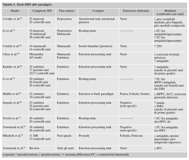
63. Cerullo MA, Eliassen JC, Smith CT, et al. Bipolar I disorder and major depressive disorder show similar brain activation during depression. Bipolar Disord 2014; 16: 703-12.
64. Chen CH, Suckling J, Lennox BR, Ooi C, Bullmore ET. A quantitative meta-analysis of fMRI studies in bipolar disorder. Bipolar Disord 2011; 13: 1-15.
65. Usnich T, Spengler S, Sajonz B, Herold D, Bauer M, Bermpohl F. Perception of social stimuli in mania: an fMRI study. Psychiatry Res 2015; 231: 71-6.
66. Mullin BC, Perlman SB, Versace A, et al. An fMRI study of attentional control in the context of emotional distracters in euthymic adults with bipolar disorder. Psychiatry Res 2012; 201: 196-205.
67. Townsend JD, Torrisi SJ, Lieberman MD, Sugar CA, Bookheimer SY, Altshuler LL. Frontal-amygdala connectivity alterations during emotion downregulation in bipolar I disorder. Biol Psychiatry 2013; 73: 127-35.
68. Kanske P, Schönfelder S, Forneck J, Wessa M. Impaired regulation of emotion: neural correlates of reappraisal and distraction in bipolar disorder and unaffected relatives. Transl Psychiatry 2015; 5: e497.
69. Sepede G, De Berardis D, Campanella D. Neural correlates of negative emotion processing in bipolar disorder. Prog Neuropsychopharmacol Biol Psychiatry 2015; 60: 1-10.
70. Cerullo MA, Fleck DE, Eliassen JC, et al. A longitudinal functional connectivity analysis of the amygdala in bipolar I disorder across mood states. Bipolar Disord 2012; 14: 175-84.
71. Townsend J, Altshuler LL. Emotion processing and regulation in bipolar disorder: a review. Bipolar Disord 2012; 14: 326-39.
72. Li M, Huang C, Deng W, et al. Contrasting and convergent patterns of amygdala connectivity in mania and depression: a resting-state study. J Affect Disord 2015; 173: 53-8.
73. Torrisi S, Moody TD, Vizueta N, et al. Differences in resting corticolimbic functional connectivity in bipolar I euthymia. Bipolar Disord 2013; 15: 156-66.
74. Li CT, Tu PC, Hsieh JC, et al. Functional dysconnection in the prefrontal-amygdala circuitry in unaffected siblings of patinetns with biopolar I disorder. Bipolar Disord 2015; 17: 626-35.
75. Dickstein DP, Leibenluft E. Emotion regulation in children and adolescents: boundaries between normalcy and bipolar disorder. Dev Psychopathol 2006; 18: 1105-31.
76. McClure-Tone EB. Socioemotional functioning in bipolar disorder versus typical development: behavioral and neural differences. Clin Psychol (New York) 2009; 16: 98-113.
77. Pavuluri MN, O’Connor MM, Harral E, Sweeney JA. Affective neural circuitry during facial emotion processing in pediatric bipolar disorder. Biol Psychiatry 2007; 62: 158-67.
78. Rich BA, Fromm SJ, Berghorst LH, et al. Neural connectivity in children with bipolar disorder: impairment in the face emotion processing circuit. J Child Psychol Psychiatry 2008; 49: 88-96.
79. Pavuluri MN, Passarotti AM, Harral EM, Sweeney JA. An fMRI study of the neural correlates of incidental versus directed emotion processing in pediatric bipolar disorder. J Am Acad Child Adolesc Psychiatry 2009; 48: 308-19.
80. Brotman MA, Rich BA, Guyer AE, et al. Amygdala activation during emotion processing of neutral faces in children with severe mood dysregulation versus ADHD or bipolar disorder. Am J Psychiatry 2010; 167: 61-9.
81. Rich BA, Vinton DT, Roberson-Nay R, et al. Limbic hyperactivation during processing of neutral facial expressions in children with bipolar disorder. Proc Natl Acad Sci U S A 2006; 103: 8900-5.
82. Kalmar JH, Wang F, Chepenik LG, et al. Relation between amygdala structure and function in adolescents with bipolar disorder. J Am Acad Child Adolesc Psychiatry 2009; 48: 636-42.
83. Passarotti AM, Sweeney JA, Pavuluri MN. Emotion processing influences working memory circuits in pediatric bipolar disorder and attention-deficit/hyperactivity disorder. J Am Acad Child Adolesc Psychiatry 2010; 49: 1064-80.
84. Haldane M, Jogia J, Cobb A, Kozuch E, Kumari V, Frangou S. Changes in brain activation during working memory and facial recognition tasks in patients with bipolar disorder with lamotrigine monotherapy. Eur Neuropsychopharmacol 2008; 18: 48-54.
85. Jogia J, Haldane M, Cobb A, Kumari V, Frangou S. Pilot investigation of the changes in cortical activation during facial affect recognition with lamotrigine monotherapy in bipolar disorder. Br J Psychiatry 2008; 192: 197-201.
86. Silverstone PH, Bell EC, Willson MC, Dave S, Wilman AH. Lithium alters brain activation in bipolar disorder in a task- and state-dependent manner: an fMRI study. Ann Gen Psychiatry 2005; 4: 14.
87. Phillips ML, Travis, MJ, Fagiolini A, Kupfer DJ. Medication effects in neuroimaging studies of bipolar disorder. Am J Psychiatry 2008; 165: 313-20.
88. Hassel S, Almeida JR, Frank E, et al. Prefrontal cortical and striatal activity to happy and fear faces in bipolar disorder is associated with comorbid substance abuse and eating disorder. J Affect Disord 2009; 118: 19-27.
89. Wildgruber D, Ackermann H, Kreifelts B, Ethofer T. Cerebral processing of linguistic and emotional prosody: fMRI studies. Prog Brain Res 2006; 156: 249-68.
90. Buchanan TW, Lutz K, Mirzazade S, et al. Recognition of emotional prosody and verbal components of spoken language: an fMRI study. Brain Res Cogn Brain Res 2000; 9: 227-38.
91. Wildgruber D, Riecker A, Hertrich I, et al. Identification of emotional intonation evaluated by fMRI. Neuroimage 2005; 24: 1233-41.
92. Kotz SA, Meyer M, Alter K, Besson M, von Cramon DY, Friederici AD. On the lateralization of emotional prosody: an event-related functional MR investigation. Brain Lang 2003; 86: 366-76.
93. Mitchell RL, Elliott R, Barry M, Cruttenden A, Woodruff PW. The neural response to emotional prosody, as revealed by functional magnetic resonance imaging. Neuropsychologia 2003; 41: 1410-21.
94. Wildgruber D, Hertrich I, Riecker A, et al. Distinct frontal regions subserve evaluation of linguistic and emotional aspects of speech intonation. Cereb Cortex 2004; 14: 1384-9.
95. Mitchell RL, Elliott R, Barry M, Cruttenden A, Woodruff PW. Neural response to emotional prosody in schizophrenia and in bipolar affective disorder. Br J Psychiatry 2004; 184: 223-30.
96. Piguet C, Fodoulian L, Aubry JM, Vuilleumier P, Houenou J. Bipolar disorder: functional neuroimaging markers in relatives. Neurosci Biobehav Rev 2015; 57: 284-96.
97. Gur RC, Erwin RJ, Gur RE, Zwil AS, Heimberg C, Kraemer HC. Facial emotion discrimination: II. Behavioral findings in depression. Psychiatry Res 1992; 42: 241-51.
98. Douglas KM, Porter RJ. Recognition of disgusted facial expressions in severe depression. Br J Psychiatry 2010; 197: 156-7.
99. McClure EB, Pope K, Hoberman AJ, Pine DS, Leibenluft E. Facial expression recognition in adolescents with mood and anxiety disorders. Am J Psychiatry 2003; 160: 1172-4.
100. Schaefer KL, Baumann J, Rich BA , Luckenbaugh DA, Zarate CA Jr. Perception of facial emotion in adults with bipolar or unipolar depression and controls. J Psychiatr Res 2010; 44: 1229-35.
101. Gray J, Venn H, Montagne B, et al. Bipolar patients show mood-congruent biases in sensitivity to facial expressions of emotion when exhibiting depressed symptoms, but not when exhibiting manic symptoms. Cogn Neuropsychiatry 2006; 11: 505-20.
102. Lembke A, Ketter TA. Impaired recognition of facial emotion in mania. Am J Psychiatry 2002; 159: 302-4.
103. Phillips ML, Swartz HA. A critical appraisal of neuroimaging studies of bipolar disorder: toward a new conceptualization of underlyng neural circuitry and roadmap for future research. Am J Psychiatry 2014; 171: 829-843.
104. Bozikas VP, Tonia T, Fokas K, Karavatos A, Kosmidis MH. Impaired emotion processing in remitted patients with bipolar disorder. J Affect Disord 2006; 91: 53-6.
105. Getz GE, Shear PK, Strakowski SM. Facial affect recognition deficits in bipolar disorder. J Int Neuropsychol Soc 2003; 9: 623-32.
106. Van Rheenen TE, Rossell SL. Phenomenological predictors of psychosocial function in bipolar disorder: Is there evidence that social cognitive and emotion regulation abnormalities contribute? Aust N Z J Psychiatry 2014; 48: 26-35.
107. Gratz K, Roemer L. Multidimensional assessment of emotion regulation and dysregulation: Development, factor structure, and initial validation of the Difficulties in Emotion Regulation Scale. J Psychopathol Behav Assess 2004; 26: 41-54.
108. Gross JJ. Handbook of emotion regulation. New York: The Guilford Press, 2011; p. 672.
109. Hajnal A, Varga E, Herold R, et al. P01-44 – Euthymic bipolar patients’ deficits in social cognition tasks. European Psychiatry 2010; 25 (Suppl 1): 264.
110. Hoertnagl CM, Muehlbacher M, Biedermann F, et al. Facial emotion recognition and its relationship to subjective and functional outcomes in remitted patients with bipolar I disorder. Bipolar Disord 2011; 13: 537-44.
111. Lahera G, Herrería E, Ruiz-Murugarren S, et al. P01-195 – Social cognition and general functioning in bipolar disorder. European Psychiatry 2009; 24 (Suppl 1): S583.
112. Martino DJ, Strejilevich SA, Fassi G, Marengo E, Igoa A. Theory of mind and facial emotion recognition in euthymic bipolar I and bipolar II disorders. Psychiatry Res 2011; 189: 379-84.
113. Phillips ML, Vieta E. Identifying functional neuroimaging biomarkers of bipolar disorder: toward DSM-V. Schizophr. Bull 2007; 33: 893-904.
114. Singh I, Rose N. Biomarkers in psychiatry. Nature 2009; 460: 202-7.
115. Semerari A, Carcione A, Dimaggio G, et al. How to evaluate metacognitive functioning in psychotherapy? The Metacognition Assessment Scale and its applications. Clin Psychol Psychother 2003; 10: 238-61.
116. Carcione A, Dimaggio G, Conti L, et al. Metacognition Assessment Scale (MAS) V.4.0.-Manual. Roma: Terzocentro, 2010.
117. Ressler KJ, Mayberg HS. Targeting abnormal neural circuits in mood and anxiety disorders: from the laboratory to the clinic. Nat Neurosci 2007;10: 1116-24.
118. Baron-Cohen S, Leslie AM, Frith U. Does the autistic child have a “theory of mind?” Cognition 1985; 21: 37-46.
119. Pincus D, Kose S, Arana A, et al. Inverse effects of oxytocin on attributing mental activity to others in depressed and healthy subjects: a double-blind placebo controlled FMRI study. Front Psychiatry 2010; 1: 134.


![]()


